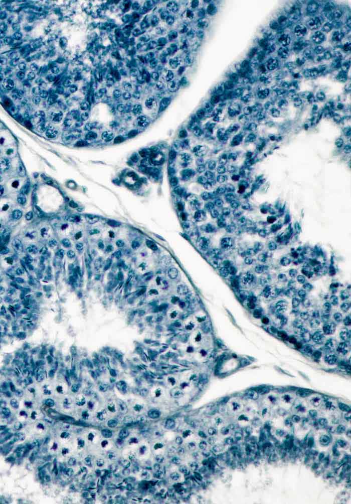
Alcian Blue pH 2.5, Umbilical Cord Histology Slides
The umbilical cord, connection between fetus and mother, is one of the most important anatomical structures related to the development of the animal during their stage of formation due to its participation as responsible of the exchange of nutrients and protection of the structural vessels.

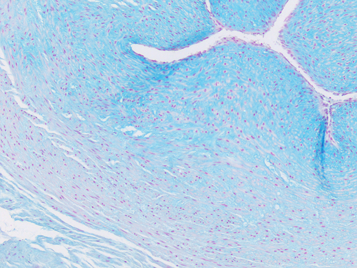
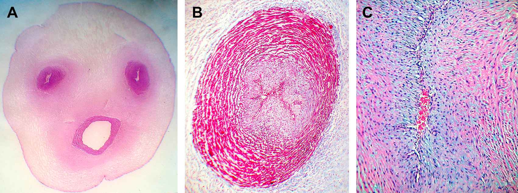
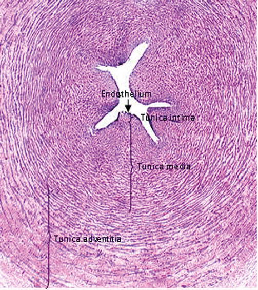
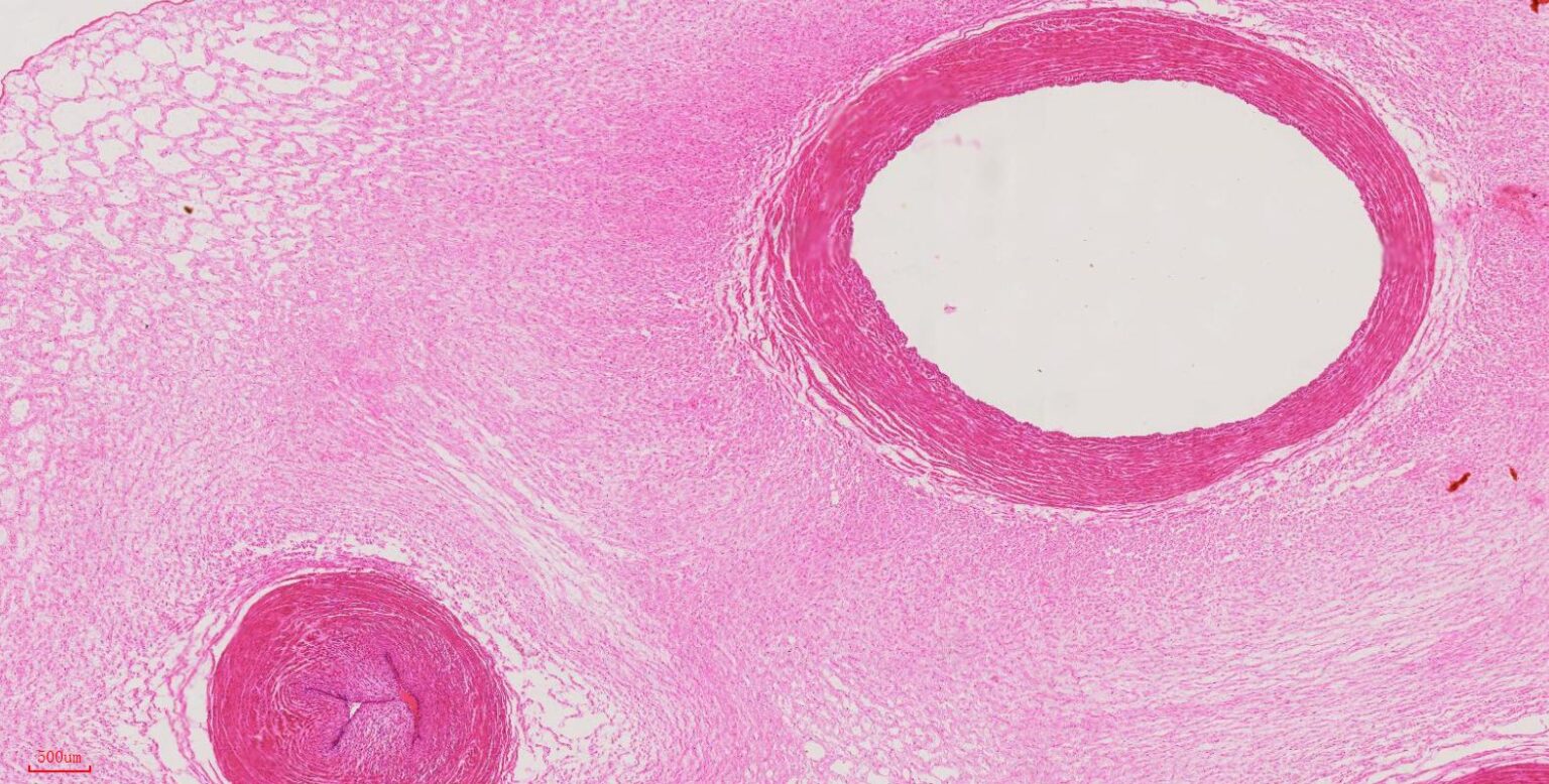
Mammal. Umbilical cord. Transverse section. 7X Mammals Mammals Reproductive system Other
1. Introduction. The human umbilical cord (hUC) is a conduit about 50-60 cm long and 1-2 cm in diameter, connecting the abdominal wall of the fetus to the placenta for exchange of nutrients and gasses during gestation [].The hUC consists of a vein and two arteries surrounded by an embryonic connective mucous tissue, known as Warton's jelly, that contains a very interesting population of.

Histologi og patologi på nett Det Medisinske Fakultet, UiO
The umbilical cord is a fetal organ connecting the placenta to the developing fetus. This structure allows for the passage of oxygen and nutrients from the maternal circulation to the fetal circulation. In doing so, it also functions to remove waste products from fetal circulation. The umbilical cord attaches to the fetus at the umbilicus and.
Alcian Blue pH 2.5, Umbilical Cord Histology Slides
HISTOLOGY, Connective tissue lab, Umbilical Index. Areolar Index:. Slide Identification: Box: Images: Umbilical cord (mucous CT) Ward's 93 W 3215 : 4: 17X Objective: 20X Objective: 20X Objective: Click on the any of the small images above to see a higher resolution image..
Histopathology and histomorphometry of umbilical cord blood vessels. Findings in normal and high
Umbilical Cord STAIN Hematoxylin & Eosin IMAGE SIZE 33,900 x 35,200 pixels 4.4 GB FILE SIZE 233 MB MAGNIFICATION 40x PIXEL SIZE 0.3171 µm SOURCE Robert L. Sorenson University of Minnesota Minneapolis, MN SETTINGS Restore Defaults Display mode Viewer options
Histology of Umbilical Cord in Mammals IntechOpen
(the list of slides in the box) 1. tendon . 2. mucous connective tissue (umbilical cord, H&E ) 3. hyaline cartilage . 4. elastic cartilage . 5. fibrocartilage . 6. compact bone (longitudinal section) 7. compact bone (transverse section). Histology and Embryology. Conditions for awarding the credits, individual study, textbooks.
Mucous tissue, Umbilical cord of Fetus t.s., 7 µm sec., H&E Stain, human histology slides
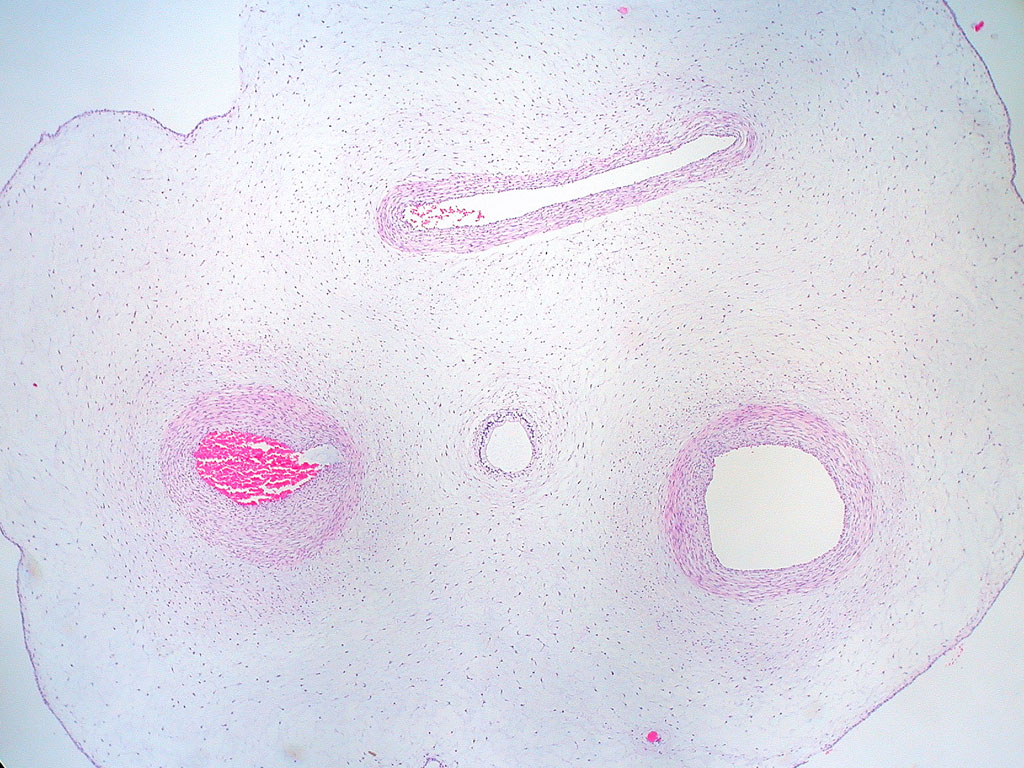
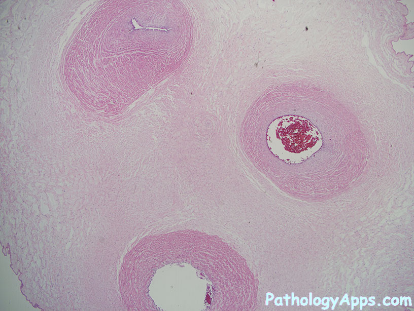
Histology @ Yale. Slide List. Umbilical Cord. Umbilical Cord The umbilical cord is mostly made up of connective tissue known as Wharton's Jelly and has relatively few cells. The cord has one large umbilical vein and two umbilical arteries. These vessels transport blood to and from the placenta, where exchange between the mother and fetus takes.
Umbilical Cord
The surface of the umbilical cord is a single layer of cuboidal to squamous amnionic epithelium. Specimen No. 32. Spleen, human, H&E. use other slides to see a more normal histology for these organs. Slide 9 notes for lymph nodes: Two lymph nodes are present on this slide. Dense connective tissue forms the capsule that surrounds the lymph.
Umbilical Cord Prepared Microscope Slide
Umbilical Cord (General Embryology) 1. UMBILICAL CORDDr. Sherif Fahmy. 2. Morphology of Umbilical Cord It is the connection between placenta and fetus. • Length: 50 - 60 cm • Diameter: 2 cm. • Shape: Tortous, showing false notes. • Contents: 2 umbilical arteries, one umbilical vein embedded in wharton's jelly and surrounded by.
cord placenta histology
HOME SLIDE BOX CHAPTER 3 - CONNECTIVE TISSUE MICROSCOPE SLIDE INFORMATION INDEX SEARCH CONTACT US TERMS OF USE SETTINGS HELP MICROSCOPE SLIDE SLIDE NAMES MH 021 Connective Tissue TISSUE Adipose (human) Areolar (human) Fascia Lata (human) Ligamentum Nuchae (cow) Umbilical Cord (human) Hematoxylin & Eosin Zenker's Formaldehyde IMAGE SIZE

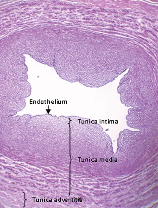
The development, structure and blood flow within the umbilical cord with particular reference to
The umbilical cord connects the fetus to the placenta, and normal function is the only mechanism for fetal oxygenation and nutrition once present.. The spirals of the cord have been studied in 5000 cords by morphometry and histology . A left twist was found in 79% with a left/right ratio of 3.7:1.0; in twins, the left direction was found in.
Histology of Umbilical Cord in Mammals IntechOpen
Pathology Outlines - Anatomy & histology-placenta & umbilical cord The placenta is a fetomaternal organ that provides gas exchange, nourishment and protection to the fetus, while the umbilical cord is the anatomic tubular structure that physically connects the developing intrauterine fetus to the placenta Menu Chapters By Subspecialty
Histology of Umbilical Cord in Mammals IntechOpen
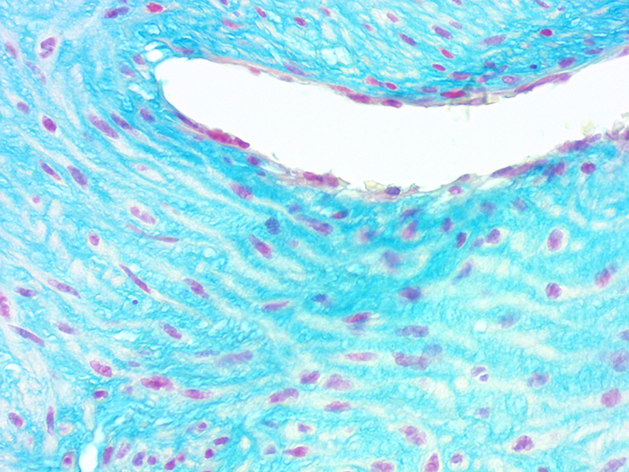
Mucoid connective tissue is a fetal tissue present in the umbilical cord. It consists of widely separated mesenchymal cells and ground substance with an abundance of hyaluronic acid. This ground substance, also referred to as Wharthon's jelly, provides insulation and protection to the blood vessels of the umbilical cord.

Mammal. Umbilical cord. Transverse section. 25X Mammals Mammals Reproductive system
The umbilical cord in humans, horses, donkeys and carnivores contains four structures: one umbilical vein, two arteries, and urachus, wrapped in mesenchymal connective tissue, also known as.

Histology Atlas Online® Umbilical Cord Slide 79
The umbilical cord is the vital connection between the fetus and the placenta. Umbilical cord development begins in the embryologic period around week 3 with the formation of the connecting stalk. By week 7, the umbilical cord has fully formed, composed of the connecting stalk, vitelline duct, and umbilical vessels surrounding the amniotic membrane. The umbilical vessels carry the fetal blood.

Histology Atlas Online® Umbilical Cord Slide 79
Describe the structural changes that occur in the uterus over the course of the menstrual cycle and pregnancy. Characterize the histological features of the oviduct, uterus, cervix, and vagina, with particular attention to the mucosal linings of these tissues.